

Cytology & Pathology
Microscopes for the pathologist and cytologist
The examination of pathology and cytology specimens places high demands on the microscopes used. Stained sections of tissue and cell compartments must be optically clearly separated from each other in order to be able to carry out a reliable assessment of structures that are rich in detail. The microscopes are often used daily for several hours at a time, and may therefore be equipped with special ergonomic features.
Cytology is a fascinating branch of biology and medicine that deals with the investigation of cells. Thanks to modern microscopes, we can take a detailed look inside the cells and observe their structures, functions and dynamics. By examining cells from various body tissues and body fluids, doctors can gain important information about the state of a patient's health. Important structures such as the cell nucleus or other cell organelles can be identified. An optical microscope offers good resolution and allows cells with a size of 0.2 to 0.3 µm to be distinguished from one another. Cytology is essential, especially in the diagnosis of cancer. Using the cytological analysis of suspect tissues, doctors can identify cancer cells and determine the severity of the tumour disease. Cytology also plays an important role in the diagnosis of infectious diseases and inflammation.
In medical science, the use of microscopes has enabled a large part of the diagnosis to be conducted on a cellular basis, together with the research and development of new drugs and treatment methods.
More advice on choosing a microscope can be found below.
Motic
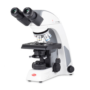
Microscope Panthera C2, bino, infinity, plan, achro, 40x-1000x, Halogen/LED
$ 2,180.00
Motic
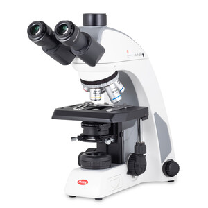
Microscope Panthera C2 Trinokular, infinity, plan, achro, 40x-1000x, 10x/22mm, Halogen/LED
$ 2,470.00
Motic
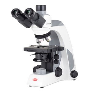
Microscope Panthera E2, Trinokular, HF, Infinity, plan achro., 40x-1000x, fixed Koehl.LED
$ 1,860.00
Euromex
DX.1153-PLi, trino microscope, 40X-1000X
$ 6,800.00
Euromex
Microscope DX.1158-PLi, trino, infinity, 10x/25, plan, 40x - 1000x, LED, 3W
$ 8,300.00
Euromex
Microscope Mikroskop DX.1158-APLi, trino, plan, apo, 40x-1000x, ergo head, AL, LED-3W
$ 11,800.00
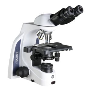
Euromex
Microscope iScope IS.1152-PLi, bino
$ 1,520.00

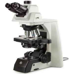
Hund
Microscope Mikroskop H 600 Wilo-Prax PL limited Edition
$ 3,050.00
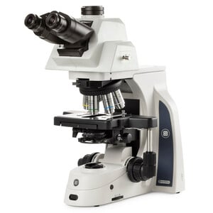
Evident Olympus
Microscope Olympus CX43 Ergo, bino, infinity, LED, w.o. objectives!
$ 5,300.00

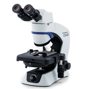
Evident Olympus
Microscope Olympus CX43 Standard, bino, LED, w.o. objectives!
$ 3,900.00
Optika
Microscope B-510ERGO, bino, ERGO, W-PLAN IOS, 40x-1000x
Our price:
RRP:
$ 2,120.00
$ 1,910.00
Optika
Microscope B-1000, Modell 1, brightfield (w.o. objectives), trino
Our price:
RRP:
$ 4,010.00
$ 3,960.00
Recommended optics
There is nothing more important in this microscope category than excellent quality optics. Two classes of objectives are best suited for the daily, demanding analysis of stained cells. On the one hand there are fluorite objectives which are marketed under their brand names such as Neofluar or Neofluor, and the even more expensive class of plan apochromat objectives.
These objectives generally have very good to excellent colour correction and field flattening, so that the user gets a true-colour representation and sees no or only minor distortions at the image edges. Both facts contribute greatly to a microscope’s high resolution.
Objective magnifications from 2x to 60x or 100x are common.
Depending on the type of specimen, objectives that have been optimised during the design process for observation with or without a coverslip are used. In this case, there are also overlaps in the areas of application, especially at smaller magnifications.
Flexibly designed versions have a correction ring that allows adjustment to the coverslip thickness up to a setting for no coverslip.
A large object field, i.e. the size of the visible image, is very important for the observation of pathological and cytological specimens. Eyepieces with a large field number ranging from 23 to even 25 are ideally suited for these tasks. The field number is printed on the eyepiece after the eyepiece magnification - for example 10x/25.
Standardised fields or scales built into the eyepiece housing allow cells to be counted and measured.
Light source
In microscopy, LED illumination is now very common. It has a comparable colour rendering index to that of natural sunlight or artificial halogen light. It also ensures an almost continuously constant colour temperature at different light intensities - unlike under a halogen light, which appears whiter the brighter it shines.
A so-called light manager has become an extremely useful feature of modern microscopes. It stores the preferred light intensity for the respective objectives, so that the user does not need to manually adjust the light intensity each time the objective is changed. In fact, objectives with higher magnification usually absorb light.
Ergonomics
What makes a microscope pleasant to use? It is features such as low-seated rotary knobs for moving the stage, or a stage that is silky-smooth in its movement. However, many pathologists also prefer to manipulate their specimens directly with their fingers. In this case, a microscope model with a very low stage may be of interest.
In the microscope head section, an ergonomically rotating tube is a great advantage in order to find the most comfortable position for microscopy. If you try it once you will never want to be without it again.