Prepared Microscope Slides
Basic component of the program are the A, B, C and D series comprising of 175 microscope slides. The four series are arranged systematically and constructively compiled, so that each enlarges the subject line of the proceeding one. They contain slides of typical micro-organisms, of cell division and of embryonic developments as well as of tissues and organs of plants, animals and man. Each of the slides has been carefully selected on the basis of its instructional value. LIEDER prepared microscope slides are made in our laboratories under scientific control. They are the product of long experience in all spheres of preparation techniques. Microtome sections are cut by highly skilled staff, cutting technique and thickness of the sections are adjusted to the objects. Out of the large number of staining techniques we select those ensuring a clear and distinct differentiation of the important structures combined with best permanency of the staining. Generally, these are complicated multicolor stainings. LIEDER prepared microscope slides are delivered on best glasses with ground edges of the size 26 x 76 mm (1 x 3"). – Every prepared microscope slide is unique and individually crafted by our well-trained technicians under rigorous scientific control. We therefore wish to point out thatdelivered products may differ from the pictures in this catalog due to natural variation of the basic raw materials and applied preparation and staining methods.
The number of series in hand should correspond approximately to the number of microscopes to allow several students to examine the same prepared microscope slides at the same time. For this reason all slides out of the series can be ordered individually also. So, important microscope slides can be supplied for all students.
SSE-30 MULTIMEDIA STUDENT PACKAGE
Typical Roots of Phanerogams, Supplementary Package of 12 items
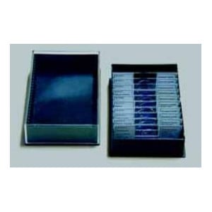
Comprising: 12 Microscope Slides in Plastic Box, Brochure with explanatory text, Cardboard box.
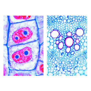
1. Herbaceous and woody roots, two t.s. on one slide
2. Young (primary) and older (secondary) roots, two t.s. on one slide
3. Salix, willow, l.s. of root showing origin of lateral roots
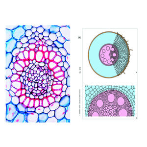
4. Iris, typical monocot root t.s. showing all structures
5. Medicago, alfalfa, root t.s. showing secondary growth
6. Tilia, lime, older woody root t.s.
7. Monstera, aerial root t.s.
8. Taraxacum, dandelion, taproot with lactiferous vessels t.s.
9. Fagus, beech, root with ectotrophic mycorrhiza, t.s.
10. Neottia nidus avis, orchid, root with endotrophic mycorrhiza, l.s.
11. Cuscuta, dodder, t.s. through stem of host showing the haustoria of the parasite
12. Pinus, older woody root t.s.
The scope of delivery and price include one or two plastic containers for 25 specimens each. Additional storage boxes can be ordered separately.