Prepared Microscope Slides
Basic component of the program are the A, B, C and D series comprising of 175 microscope slides. The four series are arranged systematically and constructively compiled, so that each enlarges the subject line of the proceeding one. They contain slides of typical micro-organisms, of cell division and of embryonic developments as well as of tissues and organs of plants, animals and man. Each of the slides has been carefully selected on the basis of its instructional value. LIEDER prepared microscope slides are made in our laboratories under scientific control. They are the product of long experience in all spheres of preparation techniques. Microtome sections are cut by highly skilled staff, cutting technique and thickness of the sections are adjusted to the objects. Out of the large number of staining techniques we select those ensuring a clear and distinct differentiation of the important structures combined with best permanency of the staining. Generally, these are complicated multicolor stainings. LIEDER prepared microscope slides are delivered on best glasses with ground edges of the size 26 x 76 mm (1 x 3"). – Every prepared microscope slide is unique and individually crafted by our well-trained technicians under rigorous scientific control. We therefore wish to point out thatdelivered products may differ from the pictures in this catalog due to natural variation of the basic raw materials and applied preparation and staining methods.
The number of series in hand should correspond approximately to the number of microscopes to allow several students to examine the same prepared microscope slides at the same time. For this reason all slides out of the series can be ordered individually also. So, important microscope slides can be supplied for all students.
Histology of domestic animals for veterinary medicine, part II
24 prepared microscope slides
- Kidney of rabbit, t.s.
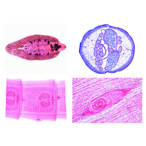
- Ovary of cat, t.s. for general study, shows primary, secondary and Graafian follicles
- Fallopian tube of pig, t.s.
- Uterus of pig, resting stage, t.s.
- Uterus of pig, pregnant stage, t.s.
- Testis and epididymis of cat, t.s.
- Sperm smear of bull
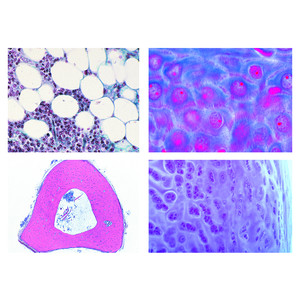
- Liver of pig, t.s.
- Brain of mouse, horizontal l.s. of the complete organ
- Cerebellum, t.s. stained by Golgi's silver method to show the Purkinje cells
- Peripheral nerve of cow or pig, l.s. routine stained
- Spinal cord of cow, t.s. stained for Nissl bodies
- Retina of pig, thin sec. special stain for details of rods and cones
- Olfactory region from nose of rabbit, t.s.
- Taste buds, t.s. of papilla foliata in tongue of rabbit shows abundant taste buds, carefully stained
- Merkel corpuscles in t.s. through snout of pig
- Hair (bristle) of pig, w.m.
- Skin of foot, cat, vertical sec. showing stratum corneum and stratum germinativum
- Mammary gland of cow, active t.s.
- Young mouse, sagittal l.s. through entire specimen passing the vertebral column
- Apis mellifica, honey bee, mouth parts of worker w.m.
- Apis mellifica, posterior leg with pollen basket w.m.
- Testis, in t.s. of abdomen of drone, Apis mellifica
- Ovary, in t.s. of abdomen of queen, Apis mellifica
The scope of delivery and price include one or two plastic containers for 25 specimens each. Additional storage boxes can be ordered separately.